FRIDAY, 25 JANUARY 2013
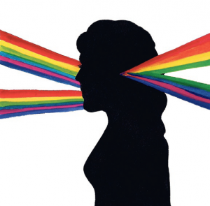
To musician edith, sound is a gastronomic experience. A major third literally tastes sweet, a perfect fourth like mown grass. But if tasting musical intervals were not strange enough, she also sees notes as colours: a C looks red, an F-sharp looks violet. Edith has a condition known as synaesthesia, which is derived from the Greek for “union of the sensations”. It means that sensations in one modality cause additional experiences in a specific second modality that was not directly stimulated.Synaesthesia has been described in the scientific literature for over 300 years. In 1689, John Locke described “a studious blind man [… who] bragged one day that he now understood what scarlet signified. [...] It was like the sound of a trumpet”. It was only in 1883 that Francis Galton investigated the experiences of synaesthetes for the first time. The majority of individuals had similar historical patterns: subjects experience synaesthesia from an early age but descriptions of such experiences were often met with ridicule, therefore discouraging self-expression, even though the experiences persisted. Within the scientific community, research into synaesthesia was neglected when the study of behaviour fell out of favour and neurophysiological studies took centre stage. The only available data on which to base the research was the subjective account of those who report their experiences. A breakthrough came in 1987, when Baron-Cohen devised a Test of Genuineness (TOG) to distinguish synaesthetes from controls based on the stable pairings of synaesthetic inducers and concurrents. Synasethetes show a 70% consistency in cross-modal percepts compared to 20-30% in controls, providing for the first time a reliable diagnosis for synaesthesia and proving its concrete existence.
Before considering the aetiology of synaesthesia, it is important to recognise this phenomenon not as a curious abnormality, but as a variant of normal human perception. A normal everyday example of such a cross-modal perception would be the perception of our body in space, where visual, somatosensory, and vestibular signals, transduced by anatomically separate sensory systems, are combined into a unitary non-ambiguous representation. Like normal perceptions, synaesthetic percepts feel real, they are not mere metaphorical comparisons of one modality with another. Perhaps because of the vividness of the percept, parallels have been drawn between synaesthesia and hallucinations, commonly experienced in psychotic conditions or under influence of psychedelic drugs like LSD. They have also been compared to visual after-images, an illusion of an image that persists after exposure to the stimulus has ceased. In imaging studies, it was found that primary sensory areas of the brain involved in colour vision are not activated in synaesthetic experiences, making the neural substrate of synaesthesia unlikely to be true colour perceptions. In keeping with the notion that synaesthesia is a variant of perception, it can manifest itself in different ways. For example, synaesthetes show a spectrum of phenotypes in the vividness of the percept: ‘projector’ synaesthetes view their percepts in the external world, e.g. they see a splash of red when hearing the note C, while ‘associators’ see them in the mind’s eye, only ‘knowing’ that C is red.
The very concept that one sensory experience can trigger another implies that sensory integration occurs at some point in the neural pathway that gives rise to the abnormal perception. The question as to how synaesthesia arises can thus be divided into two parts: what are the differences in the brain that lead to synaesthesia, and how do these differences arise? Differences that lead to abnormal sensory integration can either be due to anatomical or functional causes – there are either aberrant physical connections in the brain, or atypical functional activity present in normal cerebral circuits.
Theories concerning the anatomical basis propose cross-connections at different levels of the neuronal signalling pathways that process sensory information. This can occur locally between areas processing different aspects of a stimulus (for example colour vs shape), at a site where modalities converge (‘multisensory nexus’), or by feedback from a higher to a lower centre of the same modality. Brain connections can be measured with a technique known as Diffusion Tensor Imaging, where detection of coherent diffusion of water molecules indicates better connectivity. Evidence thus far seems to support that synaesthesia is, at least partially, caused by increased anatomical connections.
An ideal example to illustrate this is the graphemecolour synaesthesia. Sites for processing grapheme (word shape) and colour lie adjacent to each other. Functional magnetic resonance imaging (fMRI) studies have shown increased connectivity between these regions in synaesthetes, suggesting involvement of local cross-connections. Simultaneously, the parietal lobe - a multisensory nexus - also shows increased cross-connectivity. Evidence for involvement of this region comes from the fact that inhibition of this area can attenuate interference of synaesthetic colour in naming the real colour of a grapheme. Theories about anatomical connections are appealing because it is easy to visualise how cross-connections at different levels in the processing stream could explain two different phenotypes of the phenomenon: higher and lower synaesthesia. For higher synaesthetes, the conceptual properties of a grapheme trigger colours, whereas for lower synaesthetes, it is the physical properties that trigger the colour. This means that a higher synaesthete would see both the digit 4 and the Roman numeral IV in the same colour, a lower synaesthete would not. Differences in anatomical connections could even explain the variations in vividness of synaesthetic percepts: projectors have been shown to have greater connectivity in the parietal lobe compared to associators.
The fact that evidence for anatomical theories is strong does not rule out the possibility that functional abnormalities may contribute to synaesthesia. Recent research proposes that consciousness exists when a threshold number of neurons process the same information. This could mean that when processing pathways are hyperactive, additional modalities that are not normally registered could become conscious, thus leading to synaesthesia. One such example is mirror-touch synaesthesia, where tactile sensations are experienced when watching another person being touched. In these individuals, “empathy neurons”, or “mirror-neurons” are hyperactivated. This might suggest that synaesthesia is not a consequence of abnormal neuronal pathways but of a malfunction in normal pathways that are common to everyone.
Despite these controversies, and as yet unexplained observations, the question remains: how does synaesthesia develop? One current hypothesis is that it has developmental origins and is due to genetic mutations, which affect neuronal pruning. This regulatory process lasts from birth to sexual maturation and fine-tunes the neuronal structure of the brain by reducing the number of neurons and synapses, thus regulating anatomical and functional connections. The idea behind this is that all babies are born synaesthetic, and that defects in neuronal pruning would lead to synaesthesia persisting into adulthood.
While the shroud of mystery that once surrounded synaesthesia may have been lifted, our understanding of synaesthesia is far from complete. Firstly, it seems far too simplistic to suggest that the diversity of synaesthetic experience can be explained by a single mechanism. On the other hand, the fact that different members of the same family can inherit different forms of synaesthesia could mean that some forms share common neurological mechanisms. Further exploration of the genetic basis of synaesthesia will clarify the different hypotheses, and promises to unravel some of the secrets of our consciousness, perception and cerebral development.
Shi Khoo is a 3rd year student in the Department of Physiology, Development and Neuroscience. Vanda Ho is in Stage 1 Clinical School


