TUESDAY, 22 MARCH 2011
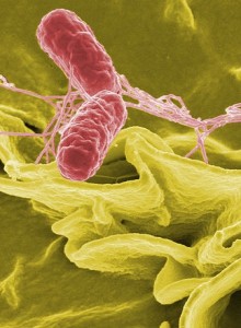
The needle complex is displayed on the outside surface of Salmonella cells and operates like a surgical needle that delivers proteins into the cells of its host, such as an infected human. These proteins can cause programmed death of the host cell or induce mechanisms that promote uptake of the bacteria.To gain insights into how this needle works, scientists in Austria have used a combination of high-resolution cryo-electron microscopy and specially-developed imaging software that allowed the complex to be modelled in fine detail [1] . Firstly, the Salmonella needles were frozen to -196 degrees Celsius so that they could be observed in almost unchanging conditions. Around 37,000 images were taken of isolated complexes, and similar images were grouped together by their newly developed image-processing algorithms using the combined power of 500 interconnected computers [2]
The result was a reconstructed Salmonella needle structure with a near-atomic resolution. Because these complexes are highly conserved among bacterial pathogens, their model could be the basis for improved understanding of the assembly and biology of this important bacterial apparatus and may help with the design of therapeutic drugs against microbial infections.
Written by Robert Jones

