SATURDAY, 19 FEBRUARY 2011
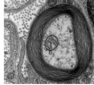
A team of scientists from the National Institute of Standards and Technology fitted a TEM with a nanofabricated 'hologram' grafting, placing it directly in the path of the spatially coherent beam of electrons. As the beam passed through the grating the electrons were diffracted. This produced multiple 'electron vortex beams'; electron beams with a corkscrew wavefront and a large orbital angular momentum. This meant that the microscope would be able to monitor how the electrons exerted torque on the subject material and how the material affected the spiral shape of the transmitted electrons, thus allowing scientists to build a more complete picture of the material’s structure.Using corksrew election beams, the distortion of the spiral wavefronts on passing though the material could be monitored, providing high-contrast, high-resolution images. This technique may find particular use in studies of biological specimens or materials with a low absorption contrast, which ordinary electrons pass through with little deflection. Although the enhanced capabilities have yet to be demonstrated, this work, published in Science, provides a significant step forwards for electron imaging [1] .
Written by Katherine Thomas

